A 17-year-old Male Patient with AML Presenting With a Suspected Fungal Infection in the ICU

Presented by Dr Rita Oladele

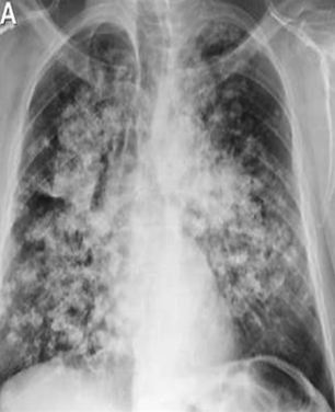
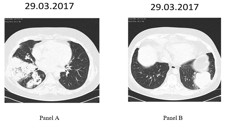
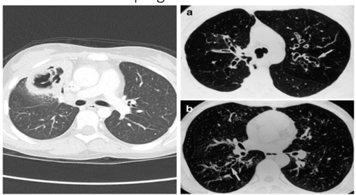
*Image is representative and is not of the actual patient.

Case Presentation

A 17-year-old male with acute myeloid leukemia (AML) presented to the emergency room 10 days post-last chemotherapy due to persistent fever (38.2°C–38.8°C), dyspnea, and cough. He had received a regular course of chemotherapy (daunorubicin, cystine arabinoside, L-asparaginase) in the previous 4 months. Physical examination revealed a body temperature of 38.4°C, blood pressure of 95/65 mm Hg, a pulse of 104 beats per minute, respiratory rate of 24 breaths per minute, and oxygen saturation of 95% (2 L O2/m was administered by nasal cannula). His initial laboratory results revealed leukocytosis (12 × 109/L, 94% neutrophils), anemia (Hemoglobin: 8.0 g/dL), and thrombocytopenia (125,000 platelets/uL). His procalcitonin was 9.1 μg/L. His chest X-ray appeared normal.

Past medical history revealed he had repeated (three) episodes of hospital-acquired infections due to an extended-spectrum beta-lactamase-producing Klebsiella pneumoniae bloodstream infection, which was managed with meropenem (2g BD 8 hourly) plus colistin (150 mg BD). He also had documented severe neutropenia (total WBC count: 500 cells/ mm3). He had been given voriconazole and trimethoprim/sulfamethoxazole as prophylaxis during earlier admissions.

He was subsequently admitted into the oncology ward and placed on voriconazole and linezolid plus piperacillin-tazobactam. A bone marrow aspirate examination was also done to assess leukemic burden, and the results revealed a morphologic leukemia-free state.

Which of the following diagnostic tests would be appropriate to evaluate a potential fungal etiology for this presentation?

  1. Serum mannan and anti-mannan testing
  2. Fungal culture, 1,3 Beta-D-Glucan (BDG), and galactomannan
  3. Candida albicans germ-tube antibody (CAGTA)
  4. All of the above

Correct answer: 2.

The most common invasive fungal infections occurring in patients in the intensive care unit (ICU) are invasive aspergillosis and invasive candidiasis, with invasive candidiasis predominating for non-neutropenic critically ill patients and invasive aspergillosis for patients with acute leukemias or receiving hematopoietic stem cell transplantation.1,2,3 In this case, with dyspnea and cough, the lungs are the most likely foci of infection. Invasive candidiasis is rarely the cause of fungal pneumonia, so a Candida-directed diagnostic strategy is not a high priority for this patient (Answer 1 mannan and anti-mannan antibodies and Answer 3 Serum CAGTA).4,5

For a suspected Aspergillus spp. infection in the ICU, the diagnosis is often driven by non-culture-based testing.1 Beta-D-Glucan (BDG), which is a pan-fungal marker and is considered appropriate for use in patients with hematologic malignancies, helps determine if a fungal cause is likely. However, it will not help determine which type of fungal infection is present. Galactomannan (GM) is appropriate for serologic testing or from bronchoalveolar lavage fluid (BAL) to help establish a probable invasive aspergillosis diagnosis. However, GM testing can yield false negatives in the setting of mold-active prophylaxis. Culture is helpful to provide support for a diagnosis of probable invasive aspergillosis if positive from sputum, BAL, bronchial brush, or aspirate.6 While imaging, such as computed tomography of the chest, is helpful for diagnosis, sometimes such testing is delayed because of cost and access issues.1 In this case, further imaging was not sought because of the negative chest X-ray.

Which of the following would be a risk factor for invasive aspergillosis in a non-neutropenic patient in the ICU?

  1. Influenza
  2. COVID-19
  3. Decompensated cirrhosis
  4. All of the above

Correct answer: 4.

While this patient has the classical hematologic malignancy factors risk for invasive aspergillosis, other patients presenting in the ICU may have a range of risk factors in the absence of neutropenia. These include influenza, COVID-19, moderate-to-severe chronic obstructive pulmonary disease, decompensated cirrhosis, uncontrolled HIV infection with CD4 count <200/mm3, and solid tumors.1 It is important to consider these risk factors and have a high index of suspicion for fungal infection in patients with pulmonary symptoms in the ICU.

Case continued

Blood cultures were repeatedly negative. Serum galactomannan and BDG were negative at this time.

On day 10 of admission, the patient was transferred to the ICU due to increasing respiratory distress (oxygen saturation of 90%) and frank hemoptysis. His lab results showed neutropenia (WBC 0.1/ mm3). A chest X-ray showed significant opacities in the lower lobes (Figure 1). As shown in Figure 2 panel A and B, a chest computer tomography (CT) scan revealed multiple nodules up to 60-mm in diameter disseminated over both lungs with ground-glass opacities. The repeat serum GM test was negative (0.2 optical density index).

Figure 1. Chest X-ray showing multiple opacities in the lower lobes of both lungs. Image courtesy of Dr Rita Oladele.


Figure 2 (Panels A and B). Two different sections of chest CTs showing multiple modules in both lungs, with several showing ground glass opacities. Images courtesy of Dr Rita Oladele.

How would you interpret the CT findings and laboratory test results?

  1. Invasive aspergillosis is unlikely in this patient because he is already on voriconazole prophylaxis.
  2. Invasive aspergillosis is unlikely in this patient due to the absence of the halo sign and the air-crescent signs.
  3. Invasive aspergillosis is unlikely because of the negative serum GM
  4. None are correct

Correct answer: 4.

The diagnosis of IPA can be difficult in ICUs and relies on integration of clinical, radiological, and microbiological results. Aspergillus mold infections can break through voriconazole prophylaxis, particularly non-fumigatus species. However, there is also a concern for Mucorales breakthrough with voriconazole prophylaxis, given the spectrum of activity of voriconazole. Mucormycosis cannot be ruled out, particularly in the setting of a negative galactomannan and BDG test results.7 The absence of typical CT findings for invasive aspergillosis does not rule out aspergillosis, particularly in the adolescent setting, where pulmonary signs are generally more non-descript.8 The finding of dense, well-circumscribed lesions is an indicative clinical feature for probable invasive pulmonary aspergillosis, with or without a halo sign.6 The negative serum galactomannan also does not rule out invasive pulmonary aspergillosis because the patient had been on mold-active antifungal prophylaxis.6 Furthermore, serum galactomannan is less sensitive than bronchoalveolar lavage for invasive aspergillosis.9

What other diagnostic tests would you recommend at this point?

  1. GM assay, microscopy, and culture of BAL
  2. Serial monitoring of serum GM test
  3. Molecular testing of serum for invasive aspergillosis
  4. None of the above

Correct answer: 1.

At this point in time, the focus of the testing should shift to the site of the infection. Continuing to test the serum is not appropriate given prior negative testing in the presence of frank pulmonary infection (answer 2). GM assay, microscopy, and culture of BAL would be helpful to establish the diagnosis.10 Molecular testing of the BAL for Aspergillus is a reasonable option here and is generally more sensitive than blood-based testing, so blood-based testing would not be recommended (answer 3).11

Case continued

Bronchoscopy and lavage revealed an erythematous airway with increased secretions, BAL was collected for GM, culture, and PCR.

  • BAL microscopy and culture were negative.
  • BAL GM was positive (Index-4.2) for Aspergillus fumigatus.
  • BAL PCR was positive (Ct-36).
  • Altered liver enzymes (AST 323 U/L, ALT 238 U/L), total bilirubin (3,1 mg/dL)
  • Repeat serial serum GM tests were 4.2, 3.8, 2.9 indices on three consecutive days.

What would be the next step(s) that should be taken in the patient's management? Commence:

  1. Itraconazole
  2. Voriconazole
  3. Liposomal amphotericin B
  4. Amphotericin B deoxycholate
  5. An echinocandin
  6. Combination therapy

Correct answer: 3

Given that the infection had already broken through azole prophylaxis and the patient’s hematologic malignancy, liposomal amphotericin was chosen. Treatment with liposomal amphotericin B was initiated with biweekly monitoring of electrolyte, urea, and creatinine levels for 3 months. A repeat CT scan showed marked improvement  (Figure 3). Liposomal amphotericin B was discontinued due to concern about renal adverse effects, and caspofungin was initiated. The duration of the treatment was seven months until total recovery, including both clinical and radiological resolution, was obtained.

Figure 3. Follow-up CTs. Three different sections of chest CTs showing general improvement of infiltrates/nodules in both lungs. Image courtesy of Dr Rita Oladele.

Resources

    1. Bassetti M, Giacobbe DR, Agvald-Ohman C, et al.; Study Group for Infections in Critically Ill Patients of the European Society of Clinical Microbiology and Infectious Diseases (ESGCIP); Fungal Infection Study Group of the European Society of Clinical Microbiology and Infectious Diseases (EFISG); European Society of Intensive Care Medicine (ESICM); European Confederation of Medical Mycology (ECMM); the Mycoses Study Group Education and Research Consortium (MSGERC), International Society of Antimicrobial Chemotherapy (ISAC); International Society for Human and Animal Mycology (ISHAM); Austrian Society for Medical Mycology (ÖGMM); Italian Society of Anesthesia, Analgesia, Reanimation, and Intensive Care (SIAARTI); Italian Society of Anti-Infective Therapy (SITA); and FUNDICU Collaborators. Invasive fungal diseases in adult patients in intensive care unit (FUNDICU): 2024 consensus definitions from ESGCIP, EFISG, ESICM, ECMM, MSGERC, ISAC, and ISHAM. Intensive Care Med. 2024;50(4):502-515. doi:10.1007/s00134-024-07341-7.
    2. Alkan A, Buyukasik Y, Uzun O, Demir AU, Coplu L. Invasive fungal infections in patients with acute leukemia: A retrospective cohort study at a tertiary-care hospital. Medicine (Baltimore). 2024;103(40):e39959. doi:10.1097/MD.0000000000039959.
    3. Kontoyiannis DP, Marr KA, Park BJ, et al. Prospective surveillance for invasive fungal infections in hematopoietic stem cell transplant recipients, 2001-2006: Overview of the Transplant-Associated Infection Surveillance Network (TRANSNET) Database. Clin Infect Dis. 2010;50(8):1091-1100. doi:10.1086/651263.
    4. Krause R, Halwachs B, Thallinger GG, et al. Characterisation of candida within the mycobiome/microbiome of the lower respiratory tract of ICU patients. PLoS One. 2016;11(5):e0155033. doi:10.1371/journal.pone.0155033. PMID: 27206014; PMCID: PMC4874575.
    5. Bassetti M, Azoulay E, Kullberg BJ, Ruhnke M, Shoham S, Vazquez J, Giacobbe DR, Calandra T. EORTC/MSGERC Definitions of invasive fungal diseases: summary of activities of the intensive care unit working group. Clin Infect Dis. 2021;72(Suppl 2):S121-S127. doi:10.1093/cid/ciaa1751.
    6. Donnelly JP, Chen SC, Kauffman CA, et al. Revision and update of the consensus definitions of invasive fungal disease from the European Organization for Research and Treatment of Cancer and the Mycoses Study Group Education and Research Consortium. Clin Infect Dis. 2020;71(6):1367-1376. doi:10.1093/cid/ciz1008.
    7. Boutin CA, Durocher F, Beauchemin S, Ziegler D, Abou Chakra CN, Dufresne SF. Breakthrough invasive fungal infections in patients with high-risk hematological disorders receiving voriconazole and posaconazole prophylaxis: a systematic review. Clin Infect Dis. 2024;79(1):151-160. doi:10.1093/cid/ciae203.
    8. Groll A, Castagnola E, Cesaro S, et al. Fourth European Conference on Infections in Leukaemia (ECIL-4): guidelines for diagnosis, prevention, and treatment of invasive fungal diseases in paediatric patients with cancer or allogeneic haemopoietic stem-cell transplantation. The Lanc Oncol. 2014;15(8). doi:10.1016/S1470-2045(14)70017-8.
    9. Wu Z, Wang L, Tan L, Wu J, Chen Z, Hu M. Diagnostic value of galactomannan in serum and bronchoalveolar lavage fluid for invasive pulmonary aspergillosis in non-neutropenic patients. Diagn Microbiol Infect Dis. 2021;99(4):115274. doi:10.1016/j.diagmicrobio.2020.115274.
    10. Hage CA, Carmona EM, Epelbaum O, Evans SE, Gabe LM, Haydour Q, Knox KS, Kolls JK, Murad MH, Wengenack NL, Limper AH. Microbiological laboratory testing in the diagnosis of fungal infections in pulmonary and critical care practice. an official American Thoracic Society clinical practice guideline. Am J Respir Crit Care Med. 2019;200(5):535-550. doi:10.1164/rccm.201906-1185ST.
    11. Imbert S, Meyer I, Palous M, Brossas JY, U et al.. AspergillusPCR in bronchoalveolar lavage fluid for the diagnosis and prognosis of aspergillosis in patients with hematological and non-hematological conditions. Front Microbiol. 2018;9:1877. doi:10.3389/fmicb.2018.01877.

Chair

Ruth Ashbee, PhD

Honorary Principal Clinical Scientist, Mycology Reference Center, Leeds, UK
Visiting Lecturer in the School of Molecular and Cellular Biology at the University of Leeds
Chair, British Society for Medical Mycology Therapeutic Drug Monitoring Guidelines Working Party
Fellow of the European Confederation of Medical Mycology
Leeds, United Kingdom

Faculty

Barbara Alexander, MD

Vice Chief of Transplant/Immunocompromised Host Infectious Diseases Services
Head of Clinical Mycology Laboratory
Professor of Medicine and Pathology
Duke University School of Medicine
Durham, North Carolina, USA

Beatriz L. Gómez, PhD

Professor, School of Medicine
Universidad del Rosario
Bogotá, Colombia

Rita Oladele, PhD

Clinical Microbiologist
Associate Professor and Clinical Consultant at University of Lagos and Lagos University Teaching Hospital
Fellow of the European Confederation of Medical Mycology
Fellow of the Royal College of Pathology
Chair of Pan Africa Mycology Working Group
Lagos, Nigeria

Joy Sarojini Michael, MD FRCPath

Professor & Clinical Microbiologist
Christian Medical College, Vellore, Tamil Nadu, India
Vice Chair of Tamil Nadu State TB Task Force Committee
Tamil Nadu, India

Alida Fe Talento, MD

Researcher and Consultant Microbiologist at Children’s Health Ireland
Clinical Senior Lecturer in the Department of Clinical Microbiology, Trinity College Dublin
Honorary Clinical Associate Professor in the Department of Microbiology at the Royal College of Surgeons
Dublin, Ireland  

Angela M. Tobón, MD

Lecturer-Investigator
Institution of Tropical Medicine
Universidad CES
Medellín, Colombia

Computer System Requirements

To participate, you will need access to a computer (either MAC or PC), tablet, or smart phone, with a Chrome (version 22.0.1229.94 or greater) web browser, Firefox (version 3.6 or greater) web browser or an Internet Explorer (version 8.0 or greater) web browser, JavaScript enabled, connected to the Internet (high speed connection preferred).

Privacy Policy

Terranova Medica, LLC, and ISHAM are committed to protecting the privacy of those who choose to participate in activities on funguseducationhub.org. The information below describes our security policies for safeguarding personal information as well as the rationale for collection and use of personal information.

Security/safeguarding personal information
This website has physical and procedural safeguards in place to prevent unauthorized access to personal information used to access this website.

Collection and use of personal information
Terranova Medica, LLC, and ISHAM do not transfer, sell, or share personal information with outside parties. We will not disclose the information unless required in a legal proceeding.

For further information, contact Tom Davis by telephone (877-276-4523), or by email (tdavis@terranovamedica.com).

Copyright © 2024 ISHAM, and Terranova Medica, LLC