24-Year-Old Man Referred to the Clinic with Severe Respiratory Symptoms, Extensive Skin Lesions, and Fever


Presented by Drs Beatriz L. Gómez, and Angela M. Tobón.

*Image is representative and is not of the actual patient.

Case Presentation

A 24-year-old male patient is referred to a regional hospital after 3 months of productive cough, dyspnea at rest, fever, anorexia, and weight loss. One month prior to consultation, he noted the appearance of non-painful skin lesions, principally on the face and extremities. He is an indigenous farmer and is a resident of a reservation in Colombia. He reports no previous diseases.

At physical examination, the patient is found to be hypotensive and tachycardic, with a respiratory rate of 22 breaths per minute and mucocutaneous pallor. Right cervical lymph node enlargement is noted. Pulmonary auscultation reveals few rhonchi in the right hemithorax. Abdomen examination shows hepatomegaly of 4 cm below the right costal margin that is painful. The neurological examination is normal. On the skin of the face and extremities are found multiple lesions in different stages of evolution: papules, vesicles, ulcers, and scabs/crusts (see Figure 1). The chest X-ray shows diffuse nodular interstitial infiltrates, no consolidation, and no pleural effusion (see chest X-ray Figure 2). Laboratory test results are shown in Table 1.

Table 1. Laboratory test results

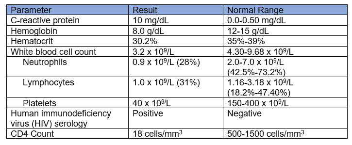
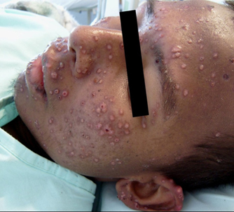
Figure 1. Skin lesions on the face and ear at different clinical stages: papules, ulcers, and scabs/crusts. Image courtesy of Angela M. Tobón.

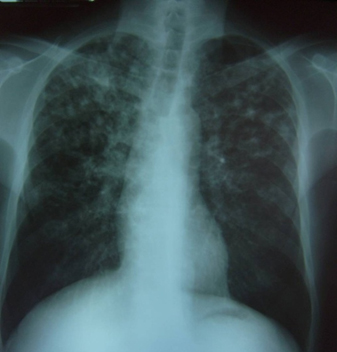
Figure 2. Chest X-ray: Bilateral nodular interstitial infiltrate, predominantly in the upper fields, no consolidation, no pleural effusion. Image courtesy of Angela M. Tobón.

What is your initial diagnostic suspicion of an opportunistic infection in this patient with advanced acquired immunodeficiency syndrome (AIDS)?

  1. Infection by Talaromyces marneffei
  2. Disseminated tuberculosis
  3. Infection by Histoplasma spp.
  4. Infection by Cryptococcus neoformans

Correct answer 3: Infection by Histoplasma spp.

Histoplasmosis, caused by Histoplasma spp., is highly endemic in the Americas.1,2 In Colombia, histoplasmosis is not a disease requiring mandatory notification, but it is known that HIV infection, especially in the AIDS stage, is present in 70% of patients diagnosed with progressive disseminated histoplasmosis (PDH).3,4 Risk factors for infection include handling soil infected with the fungus, as in the case of this patient, a farmer.5,6,7

In the clinical presentation, it is important to note the fever, constitutional and respiratory symptoms, accompanied by skin lesions consistent with disseminated histoplasmosis, which are similar to the presentation described in more than 38% of patients with AIDS.4, 5 The risk of histoplasmosis is greatest in patients with HIV infection, especially those with CD4 counts ≤200 cells/mm3.6,8,9

On the other hand, the disease produced by Talaromyces marneffei is also frequent in patients with AIDS, but it is endemic in Southeast Asia, with limited geographic distribution. In its clinical presentation it is very similar to PDH, including skin lesions in 60-70% of patients.10 In disseminated tuberculosis, skin involvement only occurs in 0.4-2.8% of patients with a predominance of respiratory symptoms.11,12  In cryptococcosis occurring in immunocompromised patients, the predominant presentation is meningeal involvement, less frequently skin involvement. When skin lesions do occur, they often appear as with an ulcerated nodule in the center, and generally, few lesions.13

Which test would you request to confirm your diagnostic suspicion?

  1. Bronchoscopy
  2. Bone marrow aspiration-biopsy
  3. Direct examination and culture of scarification of skin lesions for fungi and Mycobacterium
  4. All of the above

Correct answer 4: All of the above, depending on the local facilities

The results/reports of the direct skin examination and bone marrow are shown in Figures 3A, B, and C.

All the procedures indicated in the above question should be carried out if possible, in order to investigate the changes seen on the chest X-ray, the pancytopenia reported by the laboratory tests, and the skin lesions.

Common reasons for needing bronchoscopy are a persistent cough, infection, or something unusual seen on a chest X-ray, principally when interstitial infiltrates are reported. Bronchoscopy can also be used to obtain samples of mucus or tissue or to remove foreign bodies or other blockages from the airways or lungs. It is a minimally invasive procedure to diagnose problems with the lungs.

Wright, Giemsa, and Grocott´s methenamine silver (GMS) stains can be used to detect yeast cells of Histoplasma spp. in tissues, blood, or bone marrow smears. These cells can also be seen in tissue sections stained with Periodic Acid Schiff (PAS) but usually not with hematoxylin and eosin (H&E).6

Figure 3

A. Bone marrow aspirate smear stained with Giemsa showing numerous yeast-like cells inside macrophages (arrows) with morphology suggestive of Histoplasma spp. Magnification 100X.

B. Skin scraping stained with Grocott´s methenamine silver stain (GMS) showing numerous blastoconidia suggestive of Histoplasma spp. Magnification 100X.

C. Bone-marrow aspirate smear stained with GMS showing numerous yeast-like cells suggestive of Histoplasma spp. Magnification 40X. Images courtesy of Beatriz L. Gómez, PhD.

Does the result of the direct examination from the skin and bone marrow samples allow identification of a specific infectious agent as cause of the opportunistic infection?

  1. Yes
  2. No

Correct answer 1. Yes

Detection of the small (2- to 4-μm), oval, budding yeasts allows a presumptive diagnosis of histoplasmosis. Organisms can be found within macrophages or free in the tissues. It is unusual to find yeast cells on cytological examination of sputum or other respiratory tract fluids. The thick-walled, narrow-based budding yeast cells causing African histoplasmosis, approximately 10 to 15 μm in diameter, are about fourfold larger than those of classical Histoplasma spp. in tissue sections and might occasionally be confused with Blastomyces spp.6

Histoplasma spp. can be isolated from samples such as sputum, bronchoalveolar lavage fluid specimens, and biopsies. Culture of clinical samples is considered the gold standard; the analytical performance of the assay can vary according to the clinical presentation. In disseminated disease, useful specimens for culture include blood, urine, lymph node, and bone marrow samples, and sensitivity varies from 74% to 95% depending on the sample available. Culture does not provide a rapid result since the fungus can take up to 4 to 6 weeks to grow.6,9

Biopsy specimens of oral, cutaneous, and gastrointestinal lesions, adrenal glands, or liver and spleen can also provide a diagnosis. Sensitivity varies according to the clinical presentation, but in disseminated disease, sensitivity is around 87%.6

In cases of cutaneous tuberculosis, the skin lesions present with few or no detectable Mycobacterium bacilli.12

Since it is necessary to confirm the diagnosis of progressive disseminated histoplasmosis (PDH), and the culture can take between 20 and 30 days, which laboratory tests could be ordered to rapidly confirm the diagnosis and start prompt and appropriate therapy?

  1. Antibodies against Histoplasma spp.
  2. Histoplasma antigen
  3. Molecular tests

Correct answer 2: Histoplasma antigen

The landscape of histoplasmosis diagnosis has changed since the introduction of antigen detection tests. Detection of Histoplasma antigen in urine specimens by a commercial Histoplasma enzyme immunoassay (EIA) has good diagnostic performance and has been extensively validated in two cohorts of people living with HIV from Guatemala and Colombia, with a sensitivity of 86% and a specificity of 94% (cutoff > 0.84 ng/mL)14  in this study as well as in many other recently published studies.15 Some cross reactivity with other dimorphic fungi, such as Paracoccidioides spp.,14 and Emergomyces africanus, has been reported.16 The test is relatively easy to perform and samples are inexpensive to obtain—and the results can be obtained on the same day, with some formats in a few hours. It is also useful in the follow-up of therapy and post-therapy.14 The performance of Histoplasma antigen tests is superior to all other diagnostic tests, with a sensitivity and specificity over 90%. Detection of Histoplasma antigen has been shown to diagnose more cases when compared to conventional methods and have an impact on patients’ survival.17

The detection of antibodies using conventional serologic methods is less sensitive when performed in immunocompromised individuals. Antibodies may be negative due to the immunocompromised status of the patient; also, antibodies may take 6-8 weeks to appear and be detected. 6,18,19

Molecular testing may be a desirable alternative for correct diagnosis, however, a lack of commercially available kits and standardized methods and the limited number of validation studies, limit their broad use in clinical microbiology laboratories. Investigators have yet to determine the optimal DNA extraction method, gene target and primers, or amplification methodology.6,18,19

Case continued

A positive Histoplasma antigen result of 64 ng/mL is reported, and after confirmation of the diagnosis of histoplasmosis, treatment with liposomal amphotericin B is started at a dose of 3 mg/kg/day, with good tolerance. After 1 week of treatment, fever, anorexia, and weight loss persisted. At this time, a report was received from the bronchoalveolar lavage obtained by bronchoscopy, reporting acid-fast bacilli (AFB) and a positive PCR for Mycobacterium tuberculosis.

Discussion

Co-infections in patients with AIDS and histoplasmosis have been reported in up to 50% of cases, and reports of co-occurrence with mycobacterial disease have ranged from 2% to 35% in Latin American countries.18,20 A frequent association is reported in endemic countries for both histoplasmosis and tuberculosis,20,21 and in Colombia, co-infection occurs in 35% of the cases.22 For patients coinfected with tuberculosis, experts recommend that tetraconjugate treatment be started with isoniazid, rifampicin, pyrazinamide, and ethambutol,23 and liposomal amphotericin B can be continued for the treatment of histoplasmosis.

Case Continued

After 3 weeks of combined antifungal and antituberculosis treatment, the patient shows major clinical improvement and also a decrease in  the Histoplasma antigen titer to 30 ng/mL. Liposomal amphotericin B was stopped and replaced by oral itraconazole.  Anti-TB treatment is continued as recommended by the international guidelines.8

Can treatment for histoplasmosis be continued with oral itraconazole (drug of choice) simultaneously with anti-tuberculosis treatment including rifampicin?

  1. Yes
  2. No

Correct answer 2: No

Itraconazole and rifampicin should not be administered simultaneously, since itraconazole is a potent inhibitor of the CYP3A4 enzyme system, which induces a large number of drug interactions. Rifampicin, being an inducer of this enzyme system, decreases the therapeutic effect of itraconazole, leading to therapeutic failure.24

According to the guidelines proposed by PAHO and WHO for diagnosing and managing disseminated histoplasmosis among people living with HIV, there are several recommendations, ideally maintaining anti-tuberculosis treatment with rifampicin.

  • Extend induction therapy with amphotericin B to 1 month and perform the recommended transition to itraconazole in the maintenance phase.8
  • If the patient presents intolerance to the treatment with amphotericin B for a month or it is not possible to give this treatment for a month, in this case amphotericin B can be replaced by another azole different from itraconazole, with similar MIC (minimum inhibitory concentration) for Histoplasma spp.6,8,25 Do not use fluconazole because of high rates of resistance.25
  • Replace rifampicin by rifabutin, which leads to less propensity for drug interactions.8
  • In cases of administering simultaneously rifampicin and itraconazole, therapeutic drug monitoring should be carried out to ensure adequate itraconazole concentrations in the blood.

When it is not possible to administer liposomal amphotericin B for prolonged periods of time and rifabutin is not available, rifampicin can be replaced by a fluoroquinolone such as moxifloxacin or levofloxacin. However, this choice prolongs the anti-TB treatment due to lack of rifampicin.26 Although the literature supporting this approach is scarce,26 it is an alternative that is being used in the treatment of patients with tuberculosis resistant to rifampicin.23

Case Finalization

For our patient, rifabutin was not available and it was difficult to maintain amphotericin B for 1 month. We decided to suspend the rifampicin and replace it with levofloxacin 750 mg twice a day. The patient improved clinically after 15 days with dual treatment and with good tolerance.

  1. Araúz AB, Papineni P. Histoplasmosis. Infect Dis Clin North Am. 2021;35(2):471-491. doi:10.1016/j.idc.2021.03.011
  2. Ashraf N, Kubat RC, Poplin V, et al. Re-drawing the maps for endemic mycoses. Mycopathologia. 2020;185(5):843-865. doi:10.1007/s11046-020-00431-2
  3. Caceres DH, Knuth M, Derado G, Lindsley MD. Diagnosis of progressive disseminated histoplasmosis in advanced HIV: a meta-analysis of assay analytical performance. J Fungi. 2019;5(3):76. doi:10.3390/jof5030076
  4. Arango M, Castañeda E, Agudelo CI, et al. Colombian Histoplasmosis Study Group. Histoplasmosis: results of the Colombian national survey, 1992-2008. Biomedica. 2011; 31(3):344-56. doi:10.1590/S0120-41572011000300007
  5. Deepe GS Jr. Histoplasma capsulatum. In: Bennett JE, Dolin R, Blaser MJ, eds. In: Mandell, Douglas and Bennett´s Principles and Practice of Infectious Diseases. 9th ed. Philadelphia, PA: Elsevier; 2020:3162-3176.
  6. Thompson GR III, Gómez BL. Histoplasma, Blastomyces, Coccidioides, Paracoccidioides and other dimorphic fungi causing systemic mycoses. Caroll KC, Pfaller MA, eds. In: Manual of Clinical Microbiology, 13th ed. Washington DC: ASM Press; 2023: Chapter 125. doi:10.1128/9781683670438
  7. Gómez Londoño LF, Pérez León LC, McEwen Ochoa JG, et al. Capacity of Histoplasma capsulatum to survive the composting process. Appl Environ Soil Sci. 2019;2019:1-9. doi:10.1155/2019/5038153
  8. World Health Organization, Pan American Health Organization. Guidelines for diagnosing and managing disseminated Histoplasmosis among people living with HIV. Washington D.C.: PAHO; April 2020. https://iris.paho.org/handle/10665.2/5230
  9. Tobón AM, Gómez BL. Pulmonary histoplasmosis. Mycopathologia. 2021,86(5):697-705. doi:10.1007/s11046-021-00588-4
  10. Hospenthal, DR. Uncommon fungi and related species. In: Mandell, Douglas and Bennett´s Principles and Practice of Infectious Diseases, 9th ed. Philadelphia PA: Elsevier; 2020:3222-3237.
  11. Adenis A, Nacher M, Hanf M, et al. Tuberculosis and histoplasmosis among human immunodeficiency virus-infected patients: a comparative study. Am J Trop Med Hyg. 2014;90(2):216-23. doi:10.4269/ajtmh.13-0084
  12. Fitzgerald DW, Sterling TR, Haas DW. Mycobacterium tuberculosis. In: Mandell, Douglas and Bennett´s Principles and Practice of Infectious Diseases, 9th ed. Philadelphia PA: Elsevier; 2020:2985-3021.
  13. Perfect JR. Cryptococcosis (Cryptococcus neoformans and Cryptococcus gattii) In: Mandell, Douglas and Bennett´s Principles and Practice of Infectious Diseases, 9th ed. Philadelphia PA: Elsevier; 2020:3146-3161.
  14. Caceres DH, Scheel CM, Tobón AM, et al. Validation of an enzyme-linked immunosorbent assay that detects Histoplasma capsulatum antigenuria in Colombian patients with AIDS for diagnosis and follow-up during therapy. Clin Vaccine Immunol. 2014;21(9):1364-8. doi:10.1128/CVI.00101-14
  15. Pasqualotto AC, Queiroz-Telles F, Chebabo A, et al. The “Histoplasmosis Porto Alegre manifesto”-Addressing disseminated histoplasmosis in AIDS. PLoS Negl Trop Dis. 2023;17(1). doi:10.1371/journal.pntd.0010960
  16. Maphanga TG, Naicker SD, Gómez BL, et al. Cross-reactivity of a Histoplasma capsulatum antigen enzyme immunoassay in urine specimens from persons with emergomycosis in South Africa. Med Mycol. 2021;59(7):672-682. doi:10.1093/mmy/myaa100
  17. Medina N, Alastruey-Izquierdo A, Bonilla O, et al. A rapid screening program for histoplasmosis, tuberculosis, and cryptococcosis reduces mortality in HIV patients from Guatemala. J Fungi. 2021; 7(4):268. doi:10.3390/jof7040268
  18. Cáceres DH, Gómez BL, Tobón ÁM, et al. Tackling histoplasmosis infection in people living with HIV from Latin America: from diagnostic strategy to public health solutions. J Fungi (Basel). 2023;9(5):558. doi:10.3390/jof9050558
  19. Caceres DH, Knuth M, Derado G, Lindsley MD. Diagnosis of progressive disseminated histoplasmosis in advanced HIV: A meta-analysis of assay analytical performance. J Fungi (Basel). 2019;5(3):76. doi:10.3390/jof5030076
  20. Caceres DH, Tobon AM, Restrepo A, Chiller T, Gomez BL. The important role of co-infections in patients with AIDS and progressive disseminated histoplasmosis (PDH): A cohort from Colombia. Med Mycol Case Rep. 2018;17(19):41-4. doi:10.1016/j.mmcr.2017.07.004
  21. Caceres DH, Valdes A. Histoplasmosis and tuberculosis co-occurrence in people with advanced HIV. J Fungi (Basel). 2019;5(3):73. doi:10.3390/jof5030073
  22. Caceres DH, Tobon AM, Cleveland AA, et al. Clinical and laboratory profile of persons living with human immunodeficiency virus/acquired immune deficiency syndrome and histoplasmosis from a Colombian hospital. Am J Trop Med Hyg. 2016;95(4):918-924. doi:10.4269/ajtmh.16-0344
  23. World Health Organization. WHO consolidated guidelines on tuberculosis: module 4: treatment: drug-susceptible tuberculosis treatment. Published May 24, 2022. ISBN 978-92-4-004813-3. https://www.who.int/publications/i/item/9789240048126
  24. Jaruratanasirikul S, Sriwiriyajan S. Effect of rifampicin on the pharmacokinetics of itraconazole in normal volunteers and AIDS patients. Eur J Clin Pharmacol. 1998;54(2):155-8. doi:10.1007/s002280050437
  25. Goughenour KD, Rappleye CA. Antifungal therapeutics for dimorphic fungal pathogens. Virulence. 2017;8(2):211-221. doi:10.1080/21505594.2016.1235653
  26. Agudelo CA, Restrepo CA, Molina DA, et al. Tuberculosis and histoplasmosis co-infection in AIDS patients. Am J Trop Med Hyg. 2012;87:1094–8. doi: 10.4269/ajtmh.2012.12-0292.

Chair

Ruth Ashbee, PhD

Honorary Principal Clinical Scientist, Mycology Reference Center, Leeds, UK
Visiting Lecturer in the School of Molecular and Cellular Biology at the University of Leeds
Chair, British Society for Medical Mycology Therapeutic Drug Monitoring Guidelines Working Party
Fellow of the European Confederation of Medical Mycology
Leeds, United Kingdom

Faculty

Barbara Alexander, MD

Vice Chief of Transplant/Immunocompromised Host Infectious Diseases Services
Head of Clinical Mycology Laboratory
Professor of Medicine and Pathology
Duke University School of Medicine
Durham, North Carolina, USA

Beatriz L. Gómez, PhD

Professor, School of Medicine
Universidad del Rosario
Bogotá, Colombia

Rita Oladele, PhD

Clinical Microbiologist
Associate Professor and Clinical Consultant at University of Lagos and Lagos University Teaching Hospital
Fellow of the European Confederation of Medical Mycology
Fellow of the Royal College of Pathology
Chair of Pan Africa Mycology Working Group
Lagos, Nigeria

Joy Sarojini Michael, MD FRCPath

Professor & Clinical Microbiologist
Christian Medical College, Vellore, Tamil Nadu, India
Vice Chair of Tamil Nadu State TB Task Force Committee
Tamil Nadu, India

Alida Fe Talento, MD

Researcher and Consultant Microbiologist at Children’s Health Ireland
Clinical Senior Lecturer in the Department of Clinical Microbiology, Trinity College Dublin
Honorary Clinical Associate Professor in the Department of Microbiology at the Royal College of Surgeons
Dublin, Ireland  

Angela M. Tobón, MD

Lecturer-Investigator
Institution of Tropical Medicine
Universidad CES
Medellín, Colombia

Computer System Requirements

To participate, you will need access to a computer (either MAC or PC), tablet, or smart phone, with a Chrome (version 22.0.1229.94 or greater) web browser, Firefox (version 3.6 or greater) web browser or an Internet Explorer (version 8.0 or greater) web browser, JavaScript enabled, connected to the Internet (high speed connection preferred).

Privacy Policy

Terranova Medica, LLC, and ISHAM are committed to protecting the privacy of those who choose to participate in activities on funguseducationhub.org. The information below describes our security policies for safeguarding personal information as well as the rationale for collection and use of personal information.

Security/safeguarding personal information
This website has physical and procedural safeguards in place to prevent unauthorized access to personal information used to access this website.

Collection and use of personal information
Terranova Medica, LLC, and ISHAM do not transfer, sell, or share personal information with outside parties. We will not disclose the information unless required in a legal proceeding.

For further information, contact Tom Davis by telephone (877-276-4523), or by email (tdavis@terranovamedica.com).

Copyright © 2024 ISHAM, and Terranova Medica, LLC