15-Year-Old Female Presents to the Emergency Department in Septic Shock

Presented by Dr Alida Fe Talento

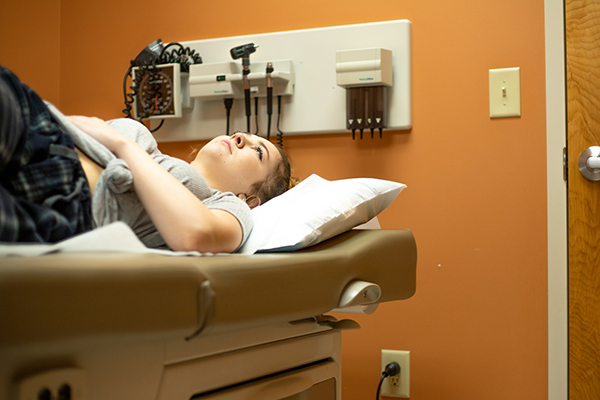
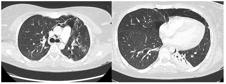
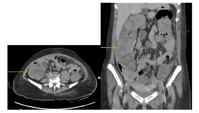
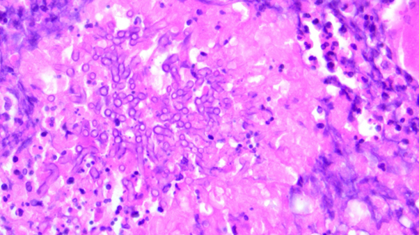
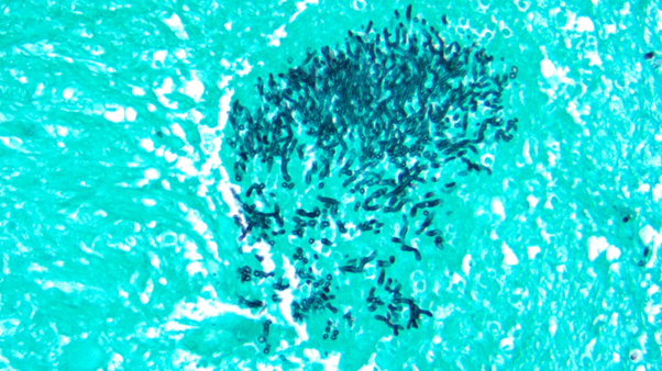
*Image is representative and is not of the actual patient.

Case Presentation

A previously well 15-year-old female presented to the emergency department (ED) with symptoms of fever, vomiting, diarrhoea, and lethargy. These symptoms were preceded by a 2-week history of an upper respiratory tract infection. On examination at the ED, she was tachycardic, hypotensive, tachypnic, and was noted to have cold, clammy, and poorly perfused extremities. The patient reported decreased urine output. Abdomen was soft and non-tender. On gynaecological examination, she was noted to have foul-smelling, green discharge per vagina. The patient was resuscitated in the ED and transferred to the paediatric ICU (PICU) for further management with a preliminary diagnosis of septic shock. She was started on broad-spectrum antimicrobial therapy (meropenem, clindamycin, vancomycin and acyclovir) after a full septic screen. She received supportive treatment in PICU, which included mechanical ventilation, renal replacement therapy, and inotropic support.

Preliminary laboratory results showed metabolic acidosis, lactic acidosis, leucocytosis with neutrophilic predominance, low haemoglobin, thrombocytopenia, and coagulopathy. Inflammatory markers, including procalcitonin, C-reactive protein, and interleukin-6, were markedly elevated. Methicillin-susceptible S. aureus was isolated from vaginal swabs. Low levels of SARS CoV-2 virus were detected on a polymerase chain reaction (PCR) test of a nasopharyngeal swab. Blood cultures were sterile. The diagnosis at this point was toxic shock syndrome due to S. aureus vs PIMS-TS (paediatric inflammatory multi-systemic syndrome following SARS-CoV-2 infection). Antimicrobial therapy was changed based on the culture results.

With supportive treatment, there were initial signs of response wherein inotropic and ventilatory support were slowly weaned. She continued to have high-grade fever and was persistently thrombocytopenic, with low haemoglobin and deranged bleeding parameters. Ferritin, fibrinogen, and creatinine kinase levels were all markedly elevated. Renal replacement therapy was resumed. Due to her persistent symptoms and laboratory results, which suggested an ongoing inflammatory process, she was referred to the Haematology Service. On her 10th hospital day, a bone marrow aspiration was performed, which revealed findings consistent with haemophagocytic lymphohistiocytosis (HLH) most likely due to the preceding S. aureus infection. Due to the above findings, she was prescribed intravenous (IV) immunoglobulin, IV pulsed methylprednisolone, and anakinra.

Which of the following is INCORRECT with regards to this patient’s risk for invasive fungal disease?

  1. Prolonged PICU care is associated with increased risk of invasive fungal disease (IFD)
  2. Treatment with anakinra alone increases a patient’s risk for IFD
  3. Prolonged antibacterial therapy is associated with increased risk of IFD
  4. There is a correlation between colonisation and infection, where colonisation with a fungal pathogen increases the patient’s risk for IFD

Best Answer: 2

Answer 2 is the best choice for an incorrect answer because the other factors listed (Answers 1,3, and 4) also increase the risk of IFD. There are several well recognised host factors that increase a patient’s risk for invasive fungal disease.1 It has been, however, increasingly recognised that patients without known “host factors” are also at risk for IFD. Patients who require prolonged ICU admission are increasingly at risk for IFD. Risk factors for this cohort of patients include prolonged antibacterial therapy, requirement for invasive devices such as intravenous catheters, mechanical ventilation, urinary catheters, and receipt of immunosuppressive therapies, to name a few. Colonisation with fungi such as Candida and Aspergillus spp have also been associated with an increased risk of IFD.2

The introduction of biologic agents in the treatment of malignant and autoimmune disorders has changed the epidemiology of IFD wherein some of the agents have been increasingly reported to increase the risk of serious fungal infections. Anakinra is an interleukin-1 receptor antagonist and is indicated for treatment of a number of autoimmune disorders. When prescribed alone, it has not been significantly associated with an increased risk of IFD. However, with prolonged treatment and administration with other biological agents and/or other immunosuppressive therapies such as steroids, anakinra has been reported to put a patient at increased risk for opportunistic infections. including IFD.3

Case continued

The patient was prescribed a prophylactic dose of liposomal amphotericin B following the hospital’s antifungal guidelines. Antibacterial therapy was reviewed and changed based on the patient’s clinical condition and laboratory results.

In the ensuing days, she remained critically unwell. Her respiratory status deteriorated, requiring re-institution of mechanical ventilation. A CTPA (Computed Tomography Pulmonary Angiography) revealed pneumomediastinum, pneumothorax, and patchy ground-glass changes on the left upper lobe (See Figure 1), which required a chest-tube insertion. Blood was noted in her airway on intubation. On her 20th hospital day, she was noted to have fresh blood per rectum. CT scan of her abdomen revealed haemoperitoneum, subsequently, half a litre of fresh blood was drained on abdominal drain insertion. At this time, she was noted to have a pressure ulcer on her back. Aspirates were collected from the endotracheal tube (ETA) as well as swabs from her pressure ulcer and vagina and were sent for culture. These samples isolated A. fumigatus (from the ETA and swab samples) and C. parapsilosis (from vaginal swab only). On the basis of these results, serum samples were collected for galactomannan (GM) and 1-3 beta-d-glucan (BDG) detection, and an EDTA sample was collected for Aspergillus PCR.

Figure 1. Patient was intubated, had a chest drain in situ for pneumothorax, pneumomediastinum, left lower lobe collapse, and patchy changes throughout both lungs including patchy ground glass opacification in the left upper lobe. Image used with permission.

Given the above results and her clinical status, the dose of liposomal amphotericin B was increased to treatment doses (3 mg/kg/day). Voriconazole was added several days later when susceptibility results showed that A. fumigatus was susceptible to azoles. BDG was elevated (> 523 pg/mL in 2 samples collected one week apart). However, the serum GM optical index was not suggestive of invasive aspergillosis (<0.5 in more than one sample collected one week apart). Aspergillus DNA was not detected on PCR.

Which of the following statements is CORRECT about fungal biomarkers?

  1. A serum galactomannan optical density index of <0.5 rules out invasive aspergillosis
  2. Serum BDG is a useful marker for the diagnosis of mucormycosis
  3. Receipt of mould-active antifungal agents reduces the sensitivity of the galactomannan assay
  4. Aspergillus PCR, if negative, rules out invasive aspergillosis

Discussion:

Best answer: 3

When compared to the body of evidence in adults, there are fewer data on the usefulness of fungal biomarkers in the diagnosis of IFD in children. Galactomannan assays, however, have been shown to perform similarly in children and in adults.4 It has been demonstrated that previous receipt of mould-active antifungal prophylaxis decreases the sensitivity of the GM assay (answer 3).5

BDG is a panfungal marker that is present in the fungal cell wall. Current international guidelines do not recommend its routine use for the diagnosis of IFD in children.4 In our case, BDG was elevated in 2 samples collected 1 week apart. However, this finding has to be interpreted in the context of the patient. It is important to note that the patient received IVIG treatment, which can result in a false-positive BDG.6 BDG is useful for the diagnosis of Candida and Aspergillus but not useful for Cryptococcus and Mucorales (Answer 2).

Molecular assays, when performed on tissue samples where moulds are seen on fungal stains, support the diagnosis of a proven IFD with the caveat that the methodology from DNA extraction to nucleic acid detection have been validated.

In effect, fungal biomarkers serve as adjunct in the diagnosis of IFD and should be interpreted with the clinical presentation and the host factors (Answer 4).1

Case continued

Supportive measures were continued and the patient appeared to improve. However, on her 34th hospital day, she was noted to have another episode of fresh bleeding per rectum. She was brought down to radiology for an urgent CT scan (Figure 2), which revealed findings suggestive of colitis and intraluminal haemorrhage.

Figure 2. Contrast-enhanced CT abdomen. Caecum and ascending colon wall are thickened, consistent with colitis. Intraluminal high density is also consistent with intraluminal haemorrhage (yellow arrows). Image used with permission.

Referring to Figures 1 and 2. With regards to radiological findings in IFD in children:

  1. The air crescent sign is commonly seen in early aspergillosis
  2. The reverse halo sign is associated with invasive aspergillosis (IA)
  3. Colitis is a common radiological finding in invasive aspergillosis
  4. Chest CT scan showing pulmonary nodules with surrounding ground glass opacity are suggestive of IA

Discussion:

Best answer: 4

The lungs are the usual portal of entry of Aspergillus spores thus most patients present with pulmonary involvement. It is important to ascertain, based on clinical presentation, if the patient has symptoms referring to other organ systems such as the central nervous system (CNS) or GI tract to ensure that the appropriate imaging techniques are requested.

Chest CT scans showing pulmonary nodules. which may be surrounded by ground-glass opacity or consolidation, are the standard findings in adult patients with pulmonary IA (Answer 4). The air crescent sign is usually a late sign of aspergillosis and may not be evident in non-neutropenic adult and paediatric patients like our case (Answer 1). Fungal pneumonia in non-neutropenic patients may also present with nonspecific infiltrates, alveolar consolidation, or ground-glass opacities with septal thickening (crazy paving). This is noted particularly in patients with preceding viral infections such as H1N1 influenzae or COVID-19.1,7 The reverse halo sign is suggestive of invasive mucormycosis (Answer 2).

Pulmonary CT findings in paediatric patients with IFD can be nondescript. Therefore, findings of any pulmonary infiltrates for a paediatric patient like this are suggestive of pulmonary IFD.4

Imaging of the GI tract may also be considered if there is involvement of the GI tract. It is important to look at involvement of the liver and spleen, particularly for patients with disseminated candidiasis. In our patient, there was evidence of colitis and intraluminal bleeding, which suggest involvement of the GI tract. Colitis can be due to multiple causes both infectious and non-infectious however in patients who are critically unwell, it is important to determine the cause to be able to administer directed therapy (Answer 3).

Other imaging modalities, such as positron emission tomography (PET)/CT, have been shown to aid in the early diagnosis of IFD and monitoring response to therapy.1,7,8

Case Continued

She was brought to theatre for an emergency laparotomy. Intraoperatively, she was noted to have adhesions most likely from previous haemorrhage and retroperitoneal perforation in the cecum with massive amount of fresh and clotted blood in the bowel lumen and peritoneum.

She underwent adhesiolysis, ileocecectomy, and stoma formation. Tissue samples were sent to histopathology. See Figures 3a and b.

Figure 3a. H and E stain. Image used with permission.

Figure 3b. Gomori-methenamine silver stain. Image used with permission.

The histopathology findings are suggestive of:

  1. Aspergillosis
  2. Candidiasis
  3. Cryptococcosis
  4. Mucormycosis

Discussion:

Best Answer: 1

Figure 3a is a Haematoxylin and Eosin (H&E) stain, while Figure 3b is a Gomori-methenamine silver stain. The figures show fungal elements within necrotising granulomas within the wall of the caecum. Septated hyphae with 45-degree branching on stains are suggestive of invasive aspergillosis (Answer 1). Images used with permission.

Case continued

The patient tolerated the procedure and was transferred back to PICU. Post-operatively, she showed clinical improvement in the ensuing days. Antibiotic therapy was discontinued. However, she continued to receive dual antifungal therapy until therapeutic drug monitoring (TDM) confirmed therapeutic levels of triazoles when liposomal amphotericin B was discontinued. Subsequently, she was transferred to the wards where she completed TDM-guided triazole treatment and was discharged home improved.

Summary

This is a very complex case of a life-threatening IFD in a young previously well patient.  Although she was previously well and an immunocompetent child, the setting of septic shock due to toxic shock syndrome, with a background of recent SARS-CoV-2 infection leading to HLH and subsequent receipt of immunosuppressive therapy with high dose corticosteroids and interleukin receptor antagonist, created a perfect storm for acquiring disseminated IFD, with involvement of her respiratory (See Figure 4) and GI tract, most likely from haematogenous dissemination of the Aspergillus spores..

Figure 4. CT thorax 2 months post initiation of antifungal therapy. A number of small pulmonary nodules are shown. One of the nodules in the left upper lobe has a faint ground glass halo (yellow arrow). Image used with permission.

The lungs are the usual portal of entry for Aspergillus spores, and extrapulmonary involvement may be seen in about 25% of cases.9 GI involvement is uncommon given that the gastric acid kills the fungal spores. This patient was receiving medications such as proton pump inhibitors to decrease gastric acidity; one can only speculate whether this contributed to the fungal dissemination to her GI tract. Additionally, it has been reported that patients with mucositis or ulcers in the GI tract can be colonised by Aspergillus spores.9

GI involvement usually leads to transmural inflammation and necrosis thus often requiring bowel resection to achieve source control, as seen in our case. A review of reported cases of aspergillosis involving the GI tract, showed that some patients did not have evidence of pulmonary disease at the time of diagnosis. and death occurred in 60% of cases.9 Although not specific for Aspergillus involvement of the GI tract, abdominal CT scan may be suggestive. The eventual value of Aspergillus serology and antigenemia has not been studied in this context. Diffuse dilatation with oedema of the bowel walls, predominantly located in the caecum, as seen in our case, with some degree of haemorrhage and necrosis. are common pathological findings.9-11

Resources

    1. Donnelly JP, Chen SC, Kauffman CA, et al. Revision and update of the consensus definitions of invasive fungal disease from the European Organization for Research and Treatment of Cancer and the Mycoses Study Group Education and Research Consortium. Clin Infect Dis. 2020;71(6):1367-1376. doi:10.1093/cid/ciz1008.
    2. Brissaud O, Guichoux J, Harambat J, et al.Invasive fungal disease in PICU: epidemiology and risk factors. Ann Intensive Care. 2012;2:6. doi:10.1186/2110-5820-2-6.
    3. Xin L, Lau SKP, and Woo PCY. Fungal infection risks associated with the use of cytokine antagonists and immune checkpoint inhibitors. Exp Biol Med (Maywood). 2020;245(12):1104-1114. doi:10.1177/1535370220939862.
    4. Groll A, Castagnola E, Cesaro S, et al. Fourth European Conference on Infections in Leukaemia (ECIL-4): guidelines for diagnosis, prevention, and treatment of invasive fungal diseases in paediatric patients with cancer or allogeneic haemopoietic stem-cell transplantation. The Lanc Oncol. 2014;15(8). doi:10.1016/S1470-2045(14)70017-8.
    5. Calle-Miguel L, García-Martínez F, Corral J, et al. Changes in the epidemiology of invasive fungal disease in a pediatric hematology and oncology unit: the relevance of breakthrough infections. BMC Infectious Diseases. 2023;23:348. doi:10.1186/s12879-023-08314-9.
    6. Theel ES, Doern CD. β-D-glucan testing is important for diagnosis of invasive fungal infections. J Clin Microbiol. 2013 Nov;51(11):3478-83. doi: 10.1128/JCM.01737-13.
    7. Lewis RE, Stanzani M, Morana G, Sassi C. Radiology-based diagnosis of fungal pulmonary infections in high-risk hematology patients: are we making progress? Curr Opin Infect Dis. 2023; 36(4):250-256. doi: 10.1097/QCO.0000000000000937.
    8. Varotto A, Marchiori E, Rubello D, et al. Radiological assessment of paediatric fungal infections: a pictorial review with focus on PET/MRI. In Vivo. 2019;33:1727-1735. doi:10.21873/invivo.11663.
    9. Eggimann P, Garbino J, Marchetti O, et al. Primary Invasive aspergillosis of the digestive tract: report of two cases and review of the literature. Infection. 2006;34:333–338. doi:10.1007/s15010-006-5660-0.
    10. Karaman I, Karaman A, Boduroğlu EC, et al. Invasive aspergillus infection localized to the gastric wall: report of a case. Surg Today. 2013;43:682-684. doi:10.1007/s00595-012-0255-0.
    11. Guglielmetti S, Guaraldi G, Cogliati M, et al. Delayed diagnosis of a diffuse invasive gastrointestinal aspergillosis in an immunocompetent patient. Case Rep Crit Care. 2020;2020:3601423. doi:10.1155/2020/3601423.

Chair

Ruth Ashbee, PhD

Honorary Principal Clinical Scientist, Mycology Reference Center, Leeds, UK
Visiting Lecturer in the School of Molecular and Cellular Biology at the University of Leeds
Chair, British Society for Medical Mycology Therapeutic Drug Monitoring Guidelines Working Party
Fellow of the European Confederation of Medical Mycology
Leeds, United Kingdom

Faculty

Barbara Alexander, MD

Vice Chief of Transplant/Immunocompromised Host Infectious Diseases Services
Head of Clinical Mycology Laboratory
Professor of Medicine and Pathology
Duke University School of Medicine
Durham, North Carolina, USA

Beatriz L. Gómez, PhD

Professor, School of Medicine
Universidad del Rosario
Bogotá, Colombia

Rita Oladele, PhD

Clinical Microbiologist
Associate Professor and Clinical Consultant at University of Lagos and Lagos University Teaching Hospital
Fellow of the European Confederation of Medical Mycology
Fellow of the Royal College of Pathology
Chair of Pan Africa Mycology Working Group
Lagos, Nigeria

Joy Sarojini Michael, MD FRCPath

Professor & Clinical Microbiologist
Christian Medical College, Vellore, Tamil Nadu, India
Vice Chair of Tamil Nadu State TB Task Force Committee
Tamil Nadu, India

Alida Fe Talento, MD

Researcher and Consultant Microbiologist at Children’s Health Ireland
Clinical Senior Lecturer in the Department of Clinical Microbiology, Trinity College Dublin
Honorary Clinical Associate Professor in the Department of Microbiology at the Royal College of Surgeons
Dublin, Ireland  

Angela M. Tobón, MD

Lecturer-Investigator
Institution of Tropical Medicine
Universidad CES
Medellín, Colombia

Computer System Requirements

To participate, you will need access to a computer (either MAC or PC), tablet, or smart phone, with a Chrome (version 22.0.1229.94 or greater) web browser, Firefox (version 3.6 or greater) web browser or an Internet Explorer (version 8.0 or greater) web browser, JavaScript enabled, connected to the Internet (high speed connection preferred).

Privacy Policy

Terranova Medica, LLC, and ISHAM are committed to protecting the privacy of those who choose to participate in activities on funguseducationhub.org. The information below describes our security policies for safeguarding personal information as well as the rationale for collection and use of personal information.

Security/safeguarding personal information
This website has physical and procedural safeguards in place to prevent unauthorized access to personal information used to access this website.

Collection and use of personal information
Terranova Medica, LLC, and ISHAM do not transfer, sell, or share personal information with outside parties. We will not disclose the information unless required in a legal proceeding.

For further information, contact Tom Davis by telephone (877-276-4523), or by email (tdavis@terranovamedica.com).

Copyright © 2024 ISHAM, and Terranova Medica, LLC